AQA A Level Biology復習筆記8.4.3 Investigating the Specificity of Restriction Enzymes

Apparatus & Techniques: Investigating the Specificity of Restriction Enzymes
- The?specificity?of?restriction enzymes?can be investigated using extracted DNA and?gel electrophoresis
- Gel electrophoresis is a technique used widely in the?analysis?of DNA. During electrophoresis, an?electric current?is used to separate the DNA molecules according to their?size / mass?and their net (overall)?charge
- The separation occurs because:
- DNA?is?negatively charged?due to the phosphate groups and so when placed in an electric field the molecules move (migrate) towards the?positive electrode
- Different sized?molecules move through the gel at?different speeds. The tiny pores in the gel result in smaller molecules moving quickly, whereas larger molecules move slowly
Separation of Restriction Fragments Using Electrophoresis
- The DNA fragments produced by restriction enzymes are known as?restriction fragments
- When a sample of extracted DNA is?digested?(hydrolysed) by restriction enzymes, a number of restriction fragments of?different lengths?are produced
- The?number?and?size?of these restriction fragments can be found using?gel electrophoresis?followed by visualisation of the DNA
- First, a restriction enzyme is added to the sample of extracted DNA, creating a sample of restriction fragments
- Next, these restriction fragments are placed in a?well?that is cut into the gel nearest to the negative electrode
- An electric current is then passed through the gel, causing the negatively charged restriction fragments to?migrate?through the pores in the gel towards the positive electrode
- The restriction fragments move through the gel at different speeds due to their different sizes (lengths)
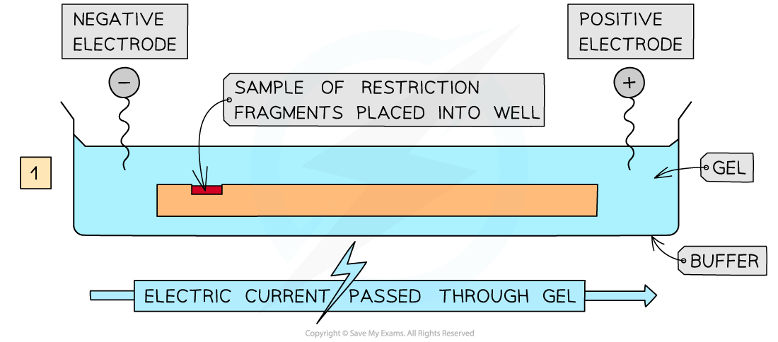
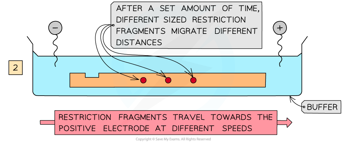
A cross-section through a gel showing how the restriction fragments migrate through it towards the positive electrode
Visualisation of Restriction Fragments
- As the restriction fragments move through the gel at different speeds due to their different sizes (lengths),?bands?of restriction fragments are formed in the gel?after electrophoresis
- However, DNA is?colourless, so the restriction fragments must be treated in such a way so that these bands can be?seen
- This can be done using a?stain, resulting in a series of coloured bands in the gel
- This can also be achieved by treating the DNA with a?radioactive marker?or adding?fluorescent?probes?that bind to the DNA
- As restriction enzymes are?highly specific, they should always produce the?same number?and?sizes?of restriction fragments (if the?same?initial sample of extracted DNA?is used)
- This means that an?unknown?restriction enzyme can be easily identified by comparing the restriction fragments it produces to those of a known restriction enzyme, as the two sets of bands produced by electrophoresis should be?exactly the same
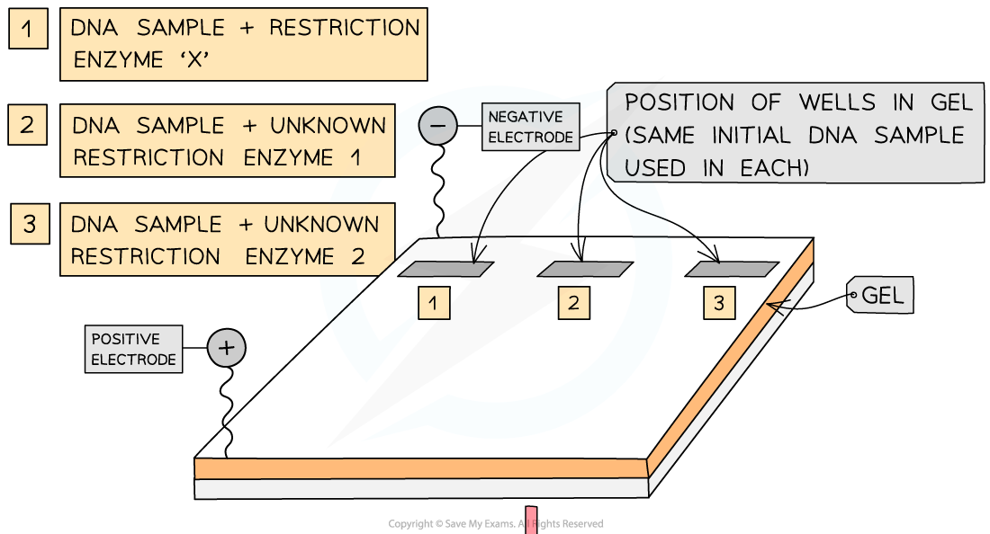
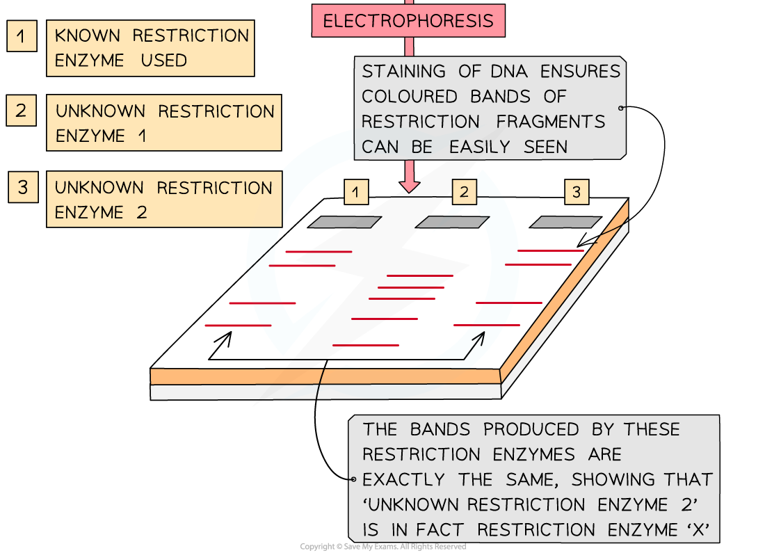
Bands of DNA (made up of the restriction fragments) are produced by electrophoresis and observed by staining the DNA. If two sets of the bands match up, then these must have been created by the same restriction enzymes, as these enzymes are highly specific.
轉載自savemyexams
以上就是關于【AQA A Level Biology復習筆記8.4.3 Investigating the Specificity of Restriction Enzymes】的解答,如需了解學校/賽事/課程動態,可至翰林教育官網獲取更多信息。
往期文章閱讀推薦:
2026 AMC10/12美國數學競賽新賽季!【翰林教育 × 清華大學出版社】獨家教材全面發售!
CAIE A-Level分數線新鮮出爐!三大考試局查分攻略+出分急救指南速覽!


翰林AMC8視頻課重磅上線!

國際競賽真題資源免費領取