OCR A Level Biology:復(fù)習(xí)筆記2.1.6 Eukaryotic Cells Under the Microscope

Photomicrographs of Eukaryotic Cells
- There are some features or structures that can help to identify whether a cell shown in an image is a plant cell or animal cell
- Structures found only in?animal?cells:?centrioles?and?microvilli
- Structures found only in?plant?cells: the?cellulose cell wall, large permanent?vacuoles?and?chloroplasts
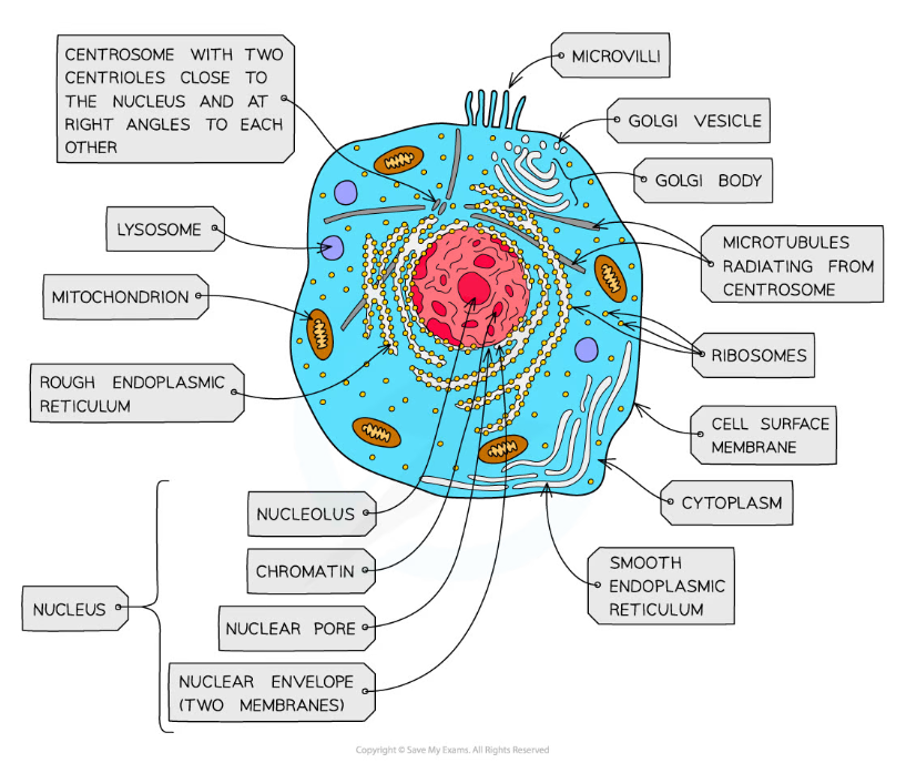
The ultrastructure of an animal cell shows a densely packed cell – the ER and RER and ribosomes form extensive networks throughout the cell in reality.
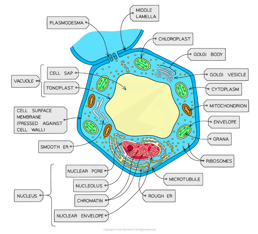
Plant cells have a larger, more regular structure in comparison to animal cells.
- Describing and interpreting photomicrographs, electron micrographs and drawings of typical animal/plant cells is an important skill
- The organelles and structures within cells have a characteristic shape and size which can be helpful when having to identify and label them in an exam
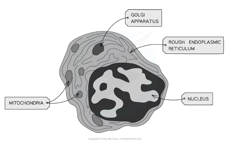
TEM electron micrograph of an animal cell showing key features. Notice the lack of a cell wall.
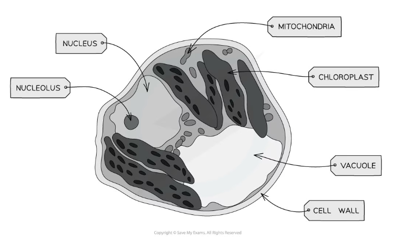
TEM electron micrograph of a plant cell showing key features. Notice the presence of a cell wall and vacuole.
- More detailed structures can be seen and identified in electron micrographs compared to photomicrographs
- This is because electron microscopes have greater maximum magnification and resolution than light (optical) microscopes
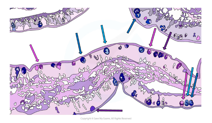
Mucus producing goblet cells (found in the lining of trachea, bronchi and larger bronchioles) are shown in a photomicrograph
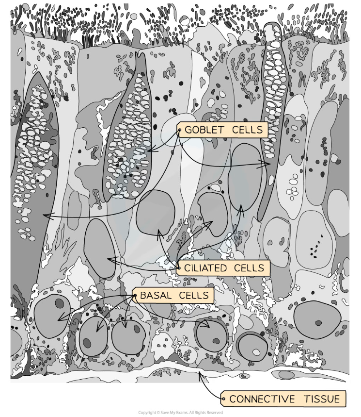
Details of the structures inside the goblet cell can be seen in an electron micrograph
Exam Tip
Make sure to learn the key identifying features of animal cells vs plant cells! It might also help to familiarise yourself with the shapes and sizes of important structures and organelles found in cells by finding some more photomicrographs and electron micrographs.
轉(zhuǎn)載自savemyexams
以上就是關(guān)于【OCR A Level Biology:復(fù)習(xí)筆記2.1.6 Eukaryotic Cells Under the Microscope】的解答,如需了解學(xué)校/賽事/課程動態(tài),可至翰林教育官網(wǎng)獲取更多信息。
往期文章閱讀推薦:
斯坦福大學(xué)官方推薦書單!名校學(xué)霸們暑假都在讀什么?
MIT官方發(fā)布【2026年夏季推薦閱讀書單】!橫跨科學(xué)/人文/經(jīng)濟(jì)...

翰林AMC8視頻課重磅上線!

國際競賽真題資源免費(fèi)領(lǐng)取