IB DP Biology: HL復習筆記3.2.5 Skills: Meiosis

Drawing the Stages of Meiosis
- Cells undergoing?meiosis?can be observed and photographed using specialized microscopes
- The different stages of meiosis have distinctive characteristics meaning they can be identified from photomicrographs
- Being able to identify the stages of meiosis from photomicrographs and diagrams is an important skill for a biologist
Step 1: Identifying if meiosis I or meiosis II is occurring
- Homologous chromosomes?pair up side by side in meiosis I only
- This means if there are?pairs of chromosomes?in a diagram or photomicrograph?meiosis I?must be occurring
- The?number of cells forming?can also help identify whether meiosis I or II is occurring
- If there are?two new cells?forming it is?meiosis I?but if there are?four new cells?forming it is?meiosis II
Identifying which stage of meiosis I is occurring
- Prophase I:?Homologous pairs?of chromosomes are visible in diploid cell (2n). Crossing over occurs
- Metaphase I: Spindle fibres pull homologous pairs so they are lined up?side by side?along the?equator?of the cell. Orientation of homologous chromosomes is random
- Anaphase I:?Whole chromosomes?are being pulled to opposite?poles?with?centromeres intact
- Telophase I:?There are?2 groups?of condensed chromosomes around which nuclei membranes are forming
- Cytokinesis:?Cytoplasm is dividing and the?cell membrane is pinching inwards?to form?two cells?with haploid chromosome numbers (n)
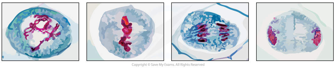
Prophase I, Metaphase I , Anaphase I and Telophase I as seen in photomicrographs
Identifying which stage of meiosis II is occurring
- Prophase II:?Single whole chromosomes?are visible in haploid cells
- Metaphase II: Single whole chromosomes are lined up along the?equator?of the cell in a?single file
- Anaphase II:?Centromeres divide?and?chromatids?are being pulled to opposite?poles
- Telophase II:?Nuclei are forming around the?4 groups?of condensed chromosomes
- Cytokinesis: Cytoplasm is dividing and?four haploid cells?are forming
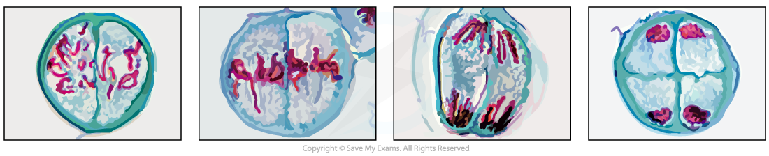
Prophase II, Metaphase II , Anaphase II and Telophase II as seen in photomicrographs
Drawing the stages of meiosis
- The distinguishing features mentioned above can also be used by biologists to draw scientific diagrams of meiosis I and meiosis II
- The conventions for drawing are:
- The drawing must have a title
- A?sharp HB pencil?should be used (and a good eraser!)
- Drawings should be on plain white paper
- Lines should be?clear,?single?lines?(no thick shading)
- No shading
- The drawing should take up as much of the space on the page as possible
- Well-defined structures should be drawn
- The drawing should be made with?proper proportions
- Label lines?should not cross or have arrowheads and should?connect directly?to the part of the drawing being labelled
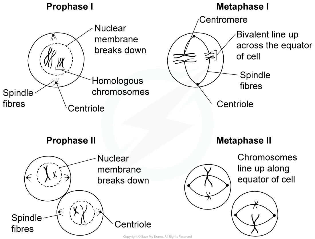
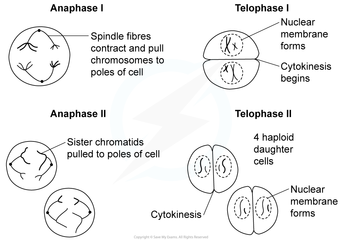
Drawing the different stages or phases of meiosis
Exam Tip
For metaphase remember?M for the middle?of the cell which is where the chromosomes will be lined up.For anaphase remember?A for away?from the middle to the poles, which is where the chromosomes / chromatids are being pulled.When drawing the stages of meiosis you do not have to show crossing over occurring.
轉載自savemyexams

國際競賽真題資源免費領取

美高學分項目重磅來襲!立即了解