AQA A Level Biology復習筆記2.2.2 Microscopy & Drawing Scientific Diagrams

Practical Skill: Microscopy & Drawing Scientific Diagrams
- Many biological structures are too small to be seen by the naked eye
- Optical microscopes are an invaluable tool for scientists as they allow for tissues, cells and organelles to be seen and studied
- For example, the movement of chromosomes during mitosis can be observed using a microscope
- When using an optical microscope always?start with the low power objective lens:
- It is?easier to find?what you are looking for in the field of view
- This helps to?prevent damage?to the lens or coverslip incase the stage has been raised too high
- ?A graticule must be used to take measurements of cells:
- A?graticule?is a small disc that has an engraved?scale.?It can be placed into the eyepiece of a microscope to act as a ruler in the field of view
- As a graticule has no fixed units it must be?calibrated?for the objective lens that is in use. This is done by using a scale engraved on a microscope slide (a stage micrometer)
- By using the two scales together the number of micrometers each graticule unit is worth can be worked out
- After this is known the graticule can be used as a?ruler?in the field of view
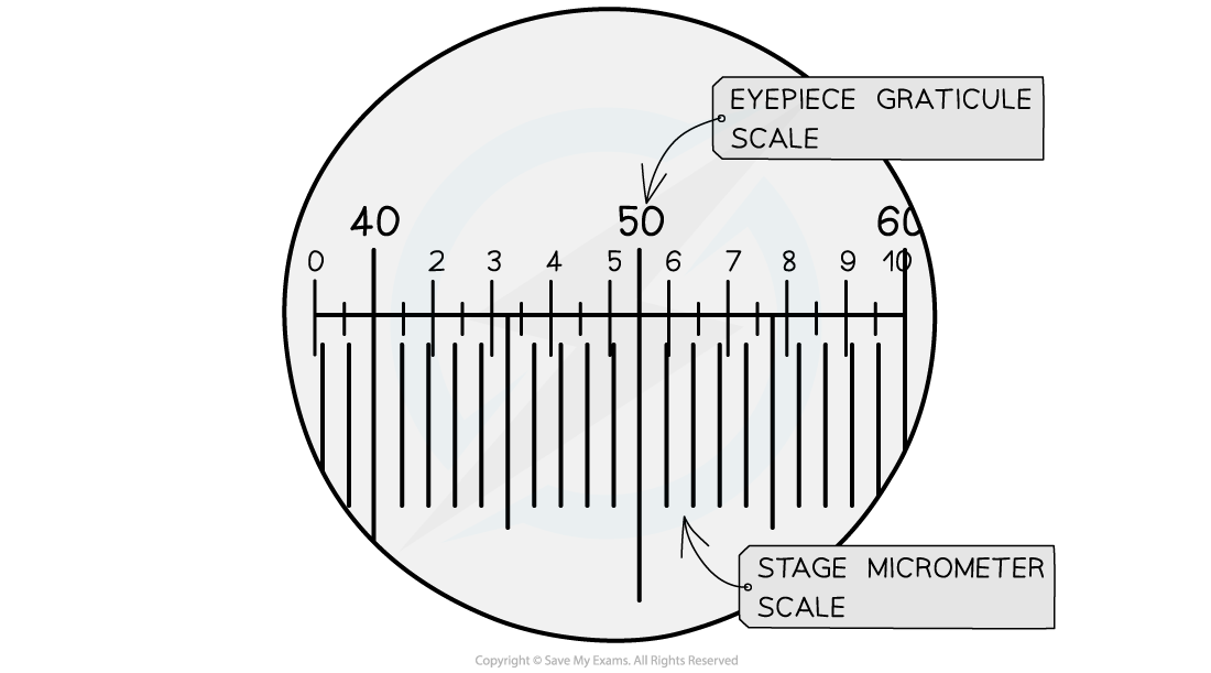
The stage micrometer scale is used to find out how many micrometers each graticule unit represents
- Electron microscopes can produce highly detailed images of animal and plant cells
- The key cellular structures within animal and plant cells are visible within the electron micrographs below
- The presence of a vacuole in a micrograph is a good indicator of the cell type
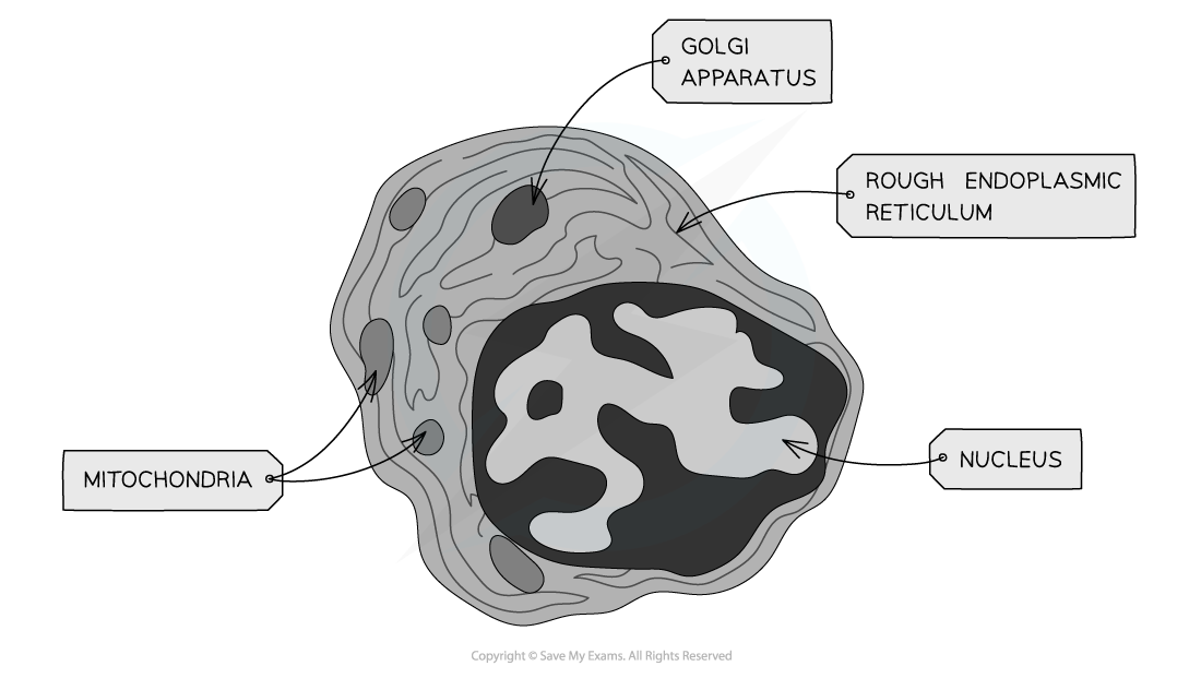
TEM electron micrograph of an animal cell showing key features
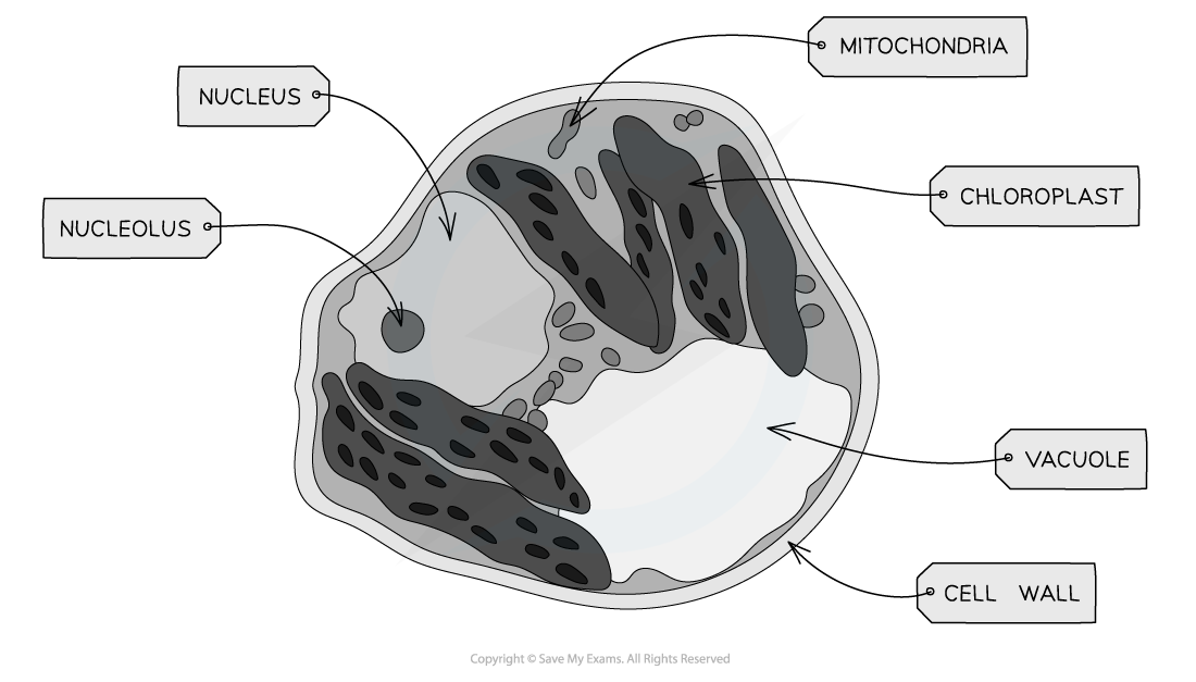
TEM electron micrograph of a plant cell showing key features
Drawing Cells
- To record the observations seen under the microscope (or from photomicrographs taken) a labelled biological drawing is often made
- Biological drawings?are line pictures which show specific features that have been observed when the specimen was viewed
- There are a number of rules/conventions that are followed when making a biological drawing
- The conventions are:
- The drawing must have a title
- The?magnification?under which the observations shown by the drawing are made must be recorded
- A?sharp HB pencil?should be used (and a good eraser!)
- Drawings should be on plain white paper
- Lines should be?clear,?single?lines?(no thick shading)
- No shading
- The drawing should take up as much of the space on the page as possible
- Well-defined structures should be drawn
- The drawing should be made with?proper proportions
- Label lines?should not cross or have arrowheads and should?connect directly?to the part of the drawing being labelled
- Label lines should be kept to one side of the drawing (in parallel to the top of the page) and drawn with a?ruler
- Drawings of cells are typically made when visualizing cells at a higher magnification power, whereas plan drawings are typically made of tissues viewed under lower magnifications (individual cells are never drawn in a plan diagram)
轉載自savemyexams
以上就是關于【AQA A Level Biology復習筆記2.2.2 Microscopy & Drawing Scientific Diagrams】的解答,如需了解學校/賽事/課程動態,可至翰林教育官網獲取更多信息。
往期文章閱讀推薦:
MIT官方發布【2026年夏季推薦閱讀書單】!橫跨科學/人文/經濟...


翰林AMC8視頻課重磅上線!

國際競賽真題資源免費領取